It uses ultrasonic sound waves in the frequency range of 1515 megahertz MHz to create images of body structures based on the pattern of echoes reflected from the tissues and organs being imaged. PRE-CLINICAL LAB ANIMALS ppt.

Increasing Ai Efficiency Using Ultrasound Ppt Download
Sonography was introduced in the Medical field in early 1950s with steady development.

. Timon travels to various vet clinics and hospitals to perform abdominal ultrasounds and echocardiograms on five or six patients like birds dogs snakes and guinea pigs a day. Drs Hertz and Edler identified heart valves and chambers 1962. An ultrasound technician may work in a clinic medical lab or hospital.
Here are some of the ins outs of the ultrasound technique including What ultrasound is how it works and what takes place during an ultrasound test. Detection of ships and mines in harbors 1946. Welcome to our newest series of articles on small animal abdominal ultrasonography.
Ultrasound imaging is a versatile well-established and widely used. B-mode US developed 1964. Ultrasound Ultrasound is defined as sound waves of frequencies greater than 20000 Hz.
Joseph Holmes brings US. Because ultrasound images are captured in real-time they can show the. A- mode developed 1948.
MedCross Imaging - ULTRASOUND - MedCross Imaging - ULTRASOUND Ultrasound imaging also called ultrasound scanning or sonography involves exposing part of the body to high-frequency sound waves to produce pictures of the inside of the body. Ultrasonography is a sound based diagnostic imaging technique used for visualising subcutaneous body structures including muscles joints vessels and internal organs for possible pathology and lesions. Furthermore therapeutic ultrasound is growing in importance to benefit animals.
Worlds Best PowerPoint Templates - CrystalGraphics offers more PowerPoint templates than anyone else in the world with over 4 million to choose from. Piezoelectric effect discovered by Paul-Jacques and Pierre Curie WWI. Frequency range of 1-10MHz max20MHz used in medical and veterinary diagnostics No disturbance to animals at employed diagnostic frequencies A sound wave travels in a pulse and when it is reflected back it becomes and echo.
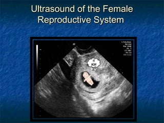
Ultrasound exams do not use ionizing radiation x-ray. The requirement of Ultrasound has gained importance in. Organs of the reproductive tract as well as a developing fetus can be viewed using ultrasound technology.
In a previous study. Ultrasound imaging encompasses a wide range of imaging modes and techniques that utilize the interaction of sound waves with living tissues to produce an image of the tissues or in the case of Doppler-based modes determine a velocity of a moving tissue primarily blood. Think horses or cattle As advancements are made in medical science ultrasound technology is becoming more and more relevant and important.
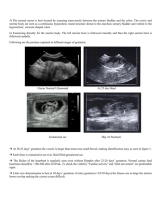
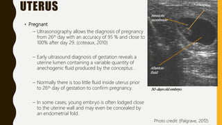
Detection of pregnancy through the use of ultrasound may be beneficial during the later stages of pregnancy day 30 or later. 2 Ultrasound timeline 1890s. The first 4 articles in the series provide an overview on the basic principles of ultrasonography while further articles will review scanning principles for each organsystem in the abdomen.
BACKGROUND The first major reference to animal testing occurred in the late nineteenth century when Louis Pasteur administered anthrax to sheep and showed the importance of vaccines with his germ theory. For example Jennifer Brooks in her Equine Wellness Magazine article Making Waves. 1983abUltrasonographic evaluation of the male genital system in small ruminants was first reported by Buckrell 1988.
The ability to identify multiple foetuses with real-time ultrasonography is a clear advantage over other techniques. Ultrasonography is the second most commonly used imaging format in veterinary practice. First use of ultrasonography in small ruminants was described in 1983 for pregnancy diagnosis both in sheep and goats by Tainturier et al.
These articles will also review the normal sonographic appearance of. Theyll give your presentations a professional memorable appearance - the kind of sophisticated look that. If they are working on animals they may work in a veterinary office a university or for a large animal care provider.
PowerPoint PPT presentation free to view ultrasound x-ray in Stafford - AV Imaging provides ultrasound services which are non-invasive imaging procedures that involve no exposure to. COMMON LABORATORY ANIMALS USED IN DRUG DEVELOPMENT. This survey assessed how veterinary point-of-care ultrasound VPOCUS including abdominal and thoracic focused assessment with sonography for trauma AFAST TFAST is used across Canada.
However unlike in the bovine application of ultrasounds in small. The best time was day 60 after matingDetermination of foetal number would allow producers to separate animals carrying singles twins or triplets for. Winner of the Standing Ovation Award for Best PowerPoint Templates from Presentations Magazine.
First time used medically in monitoring fetal development 1951. PowerPoint Presentation Last modified by. Table 1 foetal number in goats was shown as detectable at day 40 post-mating.

Small Animal Abdominal Ultrasonography Part 1 A Tour Of The Abdomen Today S Veterinary Practice

Reproductive Ultrasonography In Animals

Pregnancy Diagnose Through Ultrasound X Ray In Veterinary Field

Reproductive Ultrasonography In Animals

Ultrasound Of The Reproductive System Stacy Fielding Ppt Video Online Download

Transrectal Ultrasonography In Reproductive Management Of Cattle

0 comments
Post a Comment